- NECK PAIN
Neck pain is a common complaint. Neck muscles can be strained from poor posture — whether it’s leaning over your computer or hunching over your workbench. Osteoarthritis also is a common cause of neck pain.
Rarely, neck pain can be a symptom of a more serious problem. Seek medical care if your neck pain is accompanied by numbness or loss of strength in your arms or hands or if you have shooting pain into your shoulder or down your arm.
Symptoms
Signs and symptoms include:
- Pain that’s often worsened by holding your head in one place for long periods, such as when driving or working at a computer
- Muscle tightness and spasms
- Decreased ability to move your head
- Headache
When to see a doctor
Most neck pain improves gradually with home treatment. If not, see your doctor.
Seek immediate care if severe neck pain results from an injury, such as a motor vehicle accident, diving accident or fall.
Contact a doctor if your neck pain:
- Is severe
- Persists for several days without relief
- Spreads down arms or legs
- Is accompanied by headache, numbness, weakness or tingling
Causes
Your neck is flexible and supports the weight of your head, so it can be vulnerable to injuries and conditions that cause pain and restrict motion. Neck pain causes include:
- Muscle strains. Overuse, such as too many hours hunched over your computer or smartphone, often triggers muscle strains. Even minor things, such as reading in bed or gritting your teeth, can strain neck muscles.
- Worn joints. Just like the other joints in your body, your neck joints tend to wear down with age. Osteoarthritis causes the cushions (cartilage) between your bones (vertebrae) to deteriorate. Your body then forms bone spurs that affect joint motion and cause pain.
- Nerve compression. Herniated disks or bone spurs in the vertebrae of your neck can press on the nerves branching out from the spinal cord.
- Injuries. Rear-end auto collisions often result in whiplash injury, which occurs when the head is jerked backward and then forward, straining the soft tissues of the neck.
- Diseases. Certain diseases, such as rheumatoid arthritis, meningitis or cancer, can cause neck pain.
Prevention
Most neck pain is associated with poor posture combined with age-related wear and tear. To help prevent neck pain, keep your head centered over your spine. Some simple changes in your daily routine may help. Consider trying to:
- Use good posture. When standing and sitting, be sure your shoulders are in a straight line over your hips and your ears are directly over your shoulders.
- Take frequent breaks. If you travel long distances or work long hours at your computer, get up, move around and stretch your neck and shoulders.
- Adjust your desk, chair and computer so that the monitor is at eye level. Knees should be slightly lower than hips. Use your chair’s armrests.
- Avoid tucking the phone between your ear and shoulder when you talk. Use a headset or speakerphone instead.
- If you smoke, quit. Smoking can put you at higher risk of developing neck pain.
- Avoid carrying heavy bags with straps over your shoulder. The weight can strain your neck.
- Sleep in a good position. Your head and neck should be aligned with your body. Use a small pillow under your neck. Try sleeping on your back with your thighs elevated on pillows, which will flatten your spinal muscles.
Diagnosis
Your doctor will take a medical history and do an exam. He or she will check for tenderness, numbness and muscle weakness, as well as see how far you can move your head forward, backward and side to side.
Imaging tests
Your doctor might order imaging tests to get a better picture of the cause of your neck pain. Examples include:
- X-rays. X-rays can reveal areas in your neck where your nerves or spinal cord might be pinched by bone spurs or other degenerative changes.
- CT scan. CT scans combine X-ray images taken from many different directions to produce detailed cross-sectional views of the internal structures of your neck.
- MRI. MRI uses radio waves and a strong magnetic field to create detailed images of bones and soft tissues, including the spinal cord and the nerves coming from the spinal cord.
It’s possible to have X-ray or MRI evidence of structural problems in your neck without having symptoms. Imaging studies are best used as an adjunct to a careful history and physical exam to determine the cause of your pain.
Other tests
- Electromyography (EMG). If your doctor suspects your neck pain might be related to a pinched nerve, he or she might suggest an EMG. It involves inserting fine needles through your skin into a muscle and performing tests to measure the speed of nerve conduction to determine whether specific nerves are functioning properly.
- Blood tests. Blood tests can sometimes provide evidence of inflammatory or infectious conditions that might be causing or contributing to your neck pain.
Treatment
The most common types of mild to moderate neck pain usually respond well to self-care within two or three weeks. If neck pain persists, your doctor might recommend other treatments.
Medications
Your doctor might prescribe stronger pain medicine than what you can get over-the-counter, as well as muscle relaxants and tricyclic antidepressants for pain relief.
Therapy
- Physical therapy. A physical therapist can teach you correct posture, alignment and neck-strengthening exercises, and can use heat, ice, electrical stimulation and other measures to help ease your pain and prevent a recurrence.
- Transcutaneous electrical nerve stimulation (TENS). Electrodes placed on your skin near the painful areas deliver tiny electrical impulses that may relieve pain.
- Traction. Traction uses weights, pulleys or an air bladder to gently stretch your neck. This therapy, under supervision of a medical professional and physical therapist, may provide relief of some neck pain, especially pain related to nerve root irritation.
- Short-term immobilization. A soft collar that supports your neck may help relieve pain by taking pressure off the structures in your neck. However, if used for more than three hours at a time or for more than one to two weeks, a collar might do more harm than good.
Surgical and other procedures
- Steroid injections. Your doctor might inject corticosteroid medications near the nerve roots, into the small facet joints in the bones of the cervical spine or into the muscles in your neck to help with pain. Numbing medications, such as lidocaine, also can be injected to relieve your neck pain.
- Surgery. Rarely needed for neck pain, surgery might be an option for relieving nerve root or spinal cord compression.
- SPINAL STENOSIS
Spinal stenosis happens when the space inside the backbone is too small. This can put pressure on the spinal cord and nerves that travel through the spine. Spinal stenosis occurs most often in the lower back and the neck.
Some people with spinal stenosis have no symptoms. Others may experience pain, tingling, numbness and muscle weakness. Symptoms can get worse over time.
The most common cause of spinal stenosis is wear-and-tear changes in the spine related to arthritis. People who have severe cases of spinal stenosis may need surgery.
Surgery can create more space inside the spine. This can ease the symptoms caused by pressure on the spinal cord or nerves. But surgery can’t cure arthritis, so arthritis pain in the spine may continue.
Symptoms
Spinal stenosis often causes no symptoms. When symptoms do occur, they start slowly and get worse over time. Symptoms depend on which part of the spine is affected.
In the lower back
Spinal stenosis in the lower back can cause pain or cramping in one or both legs. This happens when you stand for a long time or when you walk. Symptoms get better when you bend forward or sit. Some people also have back pain.
In the neck
Spinal stenosis in the neck can cause:
- Numbness
- Tingling or weakness in a hand, leg, foot or arm
- Problems with walking and balance
- Neck pain
- Problems with the bowel or bladder
Causes
Spinal bones are stacked in a column from the skull to the tailbone. They protect the spinal cord, which runs through an opening called the spinal canal.
Some people are born with a small spinal canal. But most spinal stenosis occurs when something happens to reduce the amount of open space within the spine. Causes of spinal stenosis include:
- Bone spurs. Wear-and-tear damage from arthritis can cause extra bone to grow on the spine. These are called bone spurs. They can push into the spinal canal. Paget’s disease also can cause extra bone to grow on the spine.
- Herniated disks. Disks are the soft cushions that act as shock absorbers between your spinal bones. If part of the disk’s soft inner material leaks out, it can press on the spinal cord or nerves.
- Thick ligaments. The strong cords that help hold the bones of your spine together can become stiff and thick over time. Thick ligaments can push into the spinal canal.
- Tumors. Rarely, tumors can form inside the spinal canal.
- Spinal injuries. Car accidents and other trauma can cause spinal bones to break or move out of place. Swelling of nearby tissue right after back surgery also can put pressure on the spinal cord or nerves.
Risk factors
Most people with spinal stenosis are over age 50. Younger people may be at higher risk of spinal stenosis if they have scoliosis or other spinal problems.
Diagnosis
Your health care provider may ask about your symptoms and medical history. You may have a physical exam. You also may need an imaging test to help find the problem.
Imaging tests
These tests may include:
- X-rays. An X-ray of the back can show bone changes that may be making the space within the spinal canal smaller. Each X-ray involves a small dose of radiation.
- Magnetic resonance imaging (MRI). An MRI uses a powerful magnet and radio waves to produce detailed images of hard and soft tissue. The test can detect damage to the disks and ligaments. It also can show tumors that may be present.
- Computerized tomography (CT). If you can’t have an MRI, you may need a CT scan. This test combines X-ray images taken from many different angles. In a CT myelogram, a contrast dye is injected to outline the spinal cord and nerves. This can show herniated disks, bone spurs and tumors.
Treatment
Treatment for spinal stenosis depends on how severe your symptoms are.
Medications
Your health care provider might prescribe:
- Nonsteroidal anti-inflammatory drugs (NSAIDs). If common pain relievers don’t provide enough relief, prescription NSAIDs might be helpful.
- Antidepressants. Nightly doses of tricyclic antidepressants, such as amitriptyline, can help ease chronic pain.
- Anti-seizure drugs. Some anti-seizure drugs, such as gabapentin (Neurontin, Gralise), are used to reduce pain caused by damaged nerves.
- Opioids. Medications such as oxycodone (Oxycontin, Roxicodone, others) and hydrocodone (Hysingla ER) can be habit-forming.
Physical therapy
A physical therapist can teach you exercises that may help:
- Build up your strength and endurance
- Maintain the flexibility and stability of your spine
- Improve your balance
Steroid shots
Your nerve roots may become irritated and swollen at the spots where they are being pinched. Injecting a steroid medication into the space around the pinched nerve may help reduce the inflammation and relieve some of the pain.
However, steroid shots may not be the best choice for spinal stenosis. Some studies have shown that combined injections of steroids and a numbing medicine relieve back pain no better than shots of numbing medicine alone.
This is important because steroids can cause serious side effects. Repeated steroid injections can weaken nearby bones, tendons and ligaments. That’s why a person often must wait many months before getting another steroid injection.
Needle procedure for thickened ligaments
Sometimes, the ligament at the back of the lumbar spine gets too thick. Needle-like tools inserted through the skin can remove some of the ligament. This can create more space in the spinal canal to reduce pressure on nerve roots. You may be given medicine to help you feel calm during the procedure. Many people can go home the same day.
Surgery
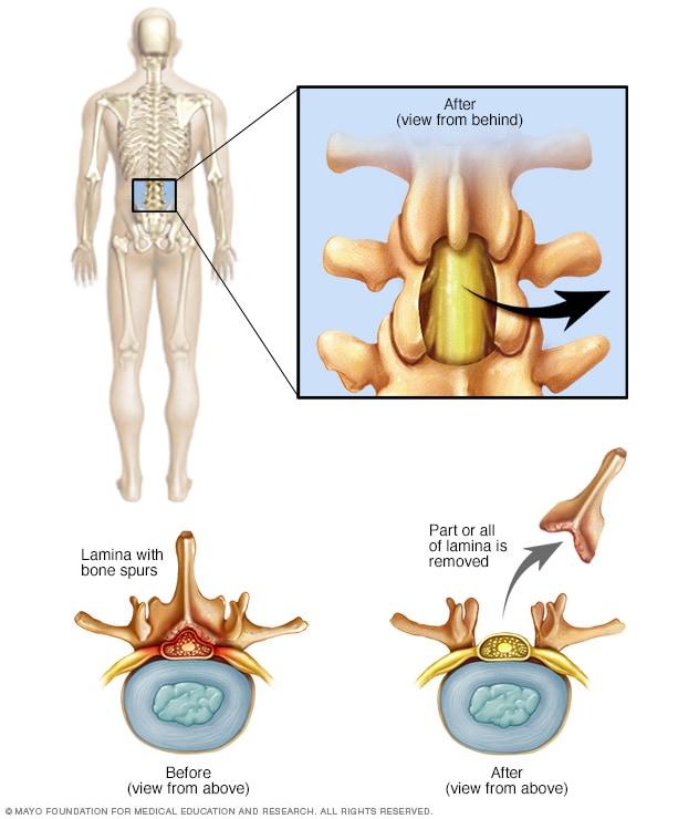
Lumbar laminectomy
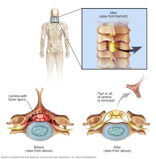
Cervical laminectomy
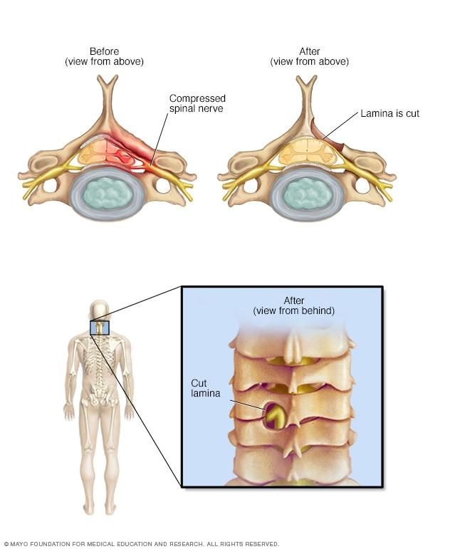
Laminotomy
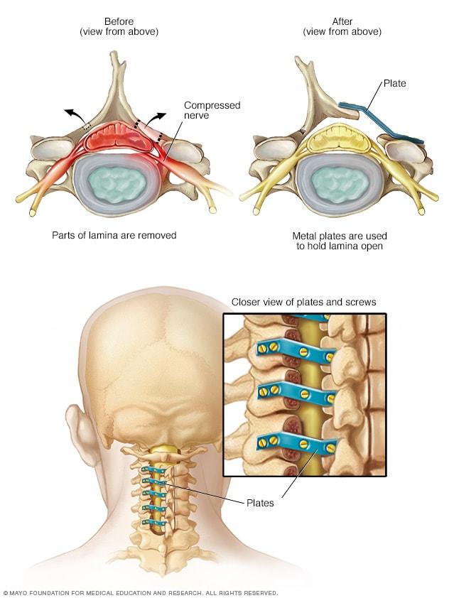
Laminoplasty
Surgeries to create more space within the spinal canal may include:
- Laminectomy. This surgery removes the back part (lamina) of the affected spinal bone. This eases pressure on the nerves by making more space around them. In some cases, that bone may need to be linked to nearby spinal bones with metal hardware and a bone graft.
- Laminotomy. This surgery removes only part of the lamina. The surgeon carves a hole just big enough to relieve pressure in a specific spot.
- Laminoplasty. This surgery is done only on spinal bones in the neck. It makes the space within the spinal canal bigger by creating a hinge on the lamina. Metal hardware bridges the gap in the opened section of the spine.
In most cases, these operations help reduce spinal stenosis symptoms. But some people’s symptoms stay the same or get worse after surgery. Surgical risks include:
- Infection
- Blood clot in a leg vein
- Tear in the membrane that covers the spinal cord
Lifestyle and home remedies
Your health care provider may suggest:
- Pain relievers.
- Weight loss. Losing excess weight can reduce pain by taking some stress off the lower back.
- Exercise. Stretching and strengthening exercises may help relieve symptoms. Talk with your health care team about what exercises are safe to do at home.
- Walking aids. In addition to providing stability, canes and walkers can help relieve pain by allowing you to bend forward while walking.